


Latest papers
See what we published
2026

Wide-field, real-time limited-angle optical diffraction tomography using massively parallel data processing
M. Sylwestrzak, W. Krauze, P. Ossowski, M. Baczewska, S. Tamborski, A. Kuś, M. Kujawińska, and M. Szkulmowski, Biomed. Opt. Express 17, 4390-4403 (2026)

Wavefront imaging with low coherence illumination simplifies the optical diffraction tomography setup and improves the signal-to-noise ratio
D.-M. Ta, M. Ziemczonok, A. Kus, P. Bon, Opt. Lett. 51, 3052-3055 (2026)

TomoTweezers: limited-angle holographic tomography realized with optical tweezers
A. Kuś, F. Dłużewski, K.Machałowski, Proc. SPIE 13857, Three-Dimensional and Multidimensional Microscopy: Image Acquisition and Processing XXXIII, 1385706 (2026);
Round robin study of Fourier ptychographic, digital holographic and hybrid holo-ptychographic microscopes
A. Kuś, M.S. Idicula, Z. Li, et al., Proc. SPIE 13861, Quantitative Phase Imaging XII, 1386104 (2026);

qtOCT: quantitative transmission optical coherence tomography
W. Krauze, M. Mazur, A. Kuś, and A. Piekarska, Biomed. Opt. Express 17, 489-498 (2026)
2025

Coded wavefront sensing for video-rate quantitative phase imaging and tomography: validation with digital holographic microscopy
S. Kazim, F. Strasser, M. Kvåle Løvmo, A. Nehrych, S. Moser, M. Ziemczonok, et al. Opt. Express 33, 25198-25209 (2025).

OSI-flex: optimization-based shearing interferometry for joint phase and shear estimation using a flexible open-source framework
J. Winnik, D. Suski, M. Heto, M. Lenarcik, M. Ziemczonok, et al., 2026 J. Phys. Photonics 8 015036

Enhanced methodology for analysis cytotoxicity of ruthenium dendrimers
A. Węgierek-Ciuk, M. Baczewska, M. Gałczyńska et al., Methods, vol. 238, p. 1-10, 2025

Tailored 3D microphantoms: An essential tool for quantitative phase tomography analysis of organoids
M. Ziemczonok, S. Desissaire, J. Neri et al., Biocybernetics and Biomedical Engineering, 2025, vol. 45, no. 2, pp. 247-257

Bio-inspired 3D-printed phantom: Encoding cellular heterogeneity for characterization of quantitative phase imaging
S. Desissaire, M. Ziemczonok, T. Cantat-Moltrecht et al., Measurement, 2025, pp. 1-10, Article no. 116765
2024
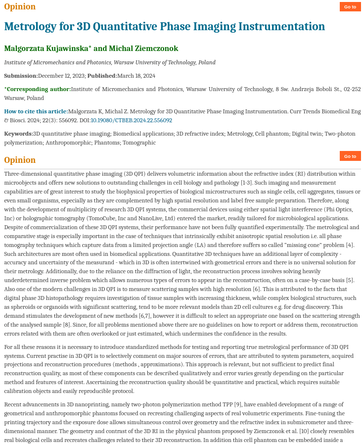
Metrology for 3D Quantitative Phase Imaging Instrumentation
M. Kujawińska, M. Ziemczonok, Current Trends in Biomedical Engineering & Biosciences, vol. 22, Art no. 4, 2024
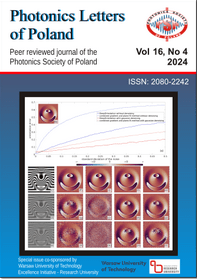
Spectral off-axis phase shifting in transmission optical coherence tomography
M. Mazur, W. Krauze, A. Kuś et al., Photonics Letters of Poland, 2024, vol. 16, no. 4, pp. 58-60
Segment and support: a dual-purpose deep learning solution for limited angle holographic tomography
M. Gontarz, W. Krauze, V. Dutta et al., Optics Express, 2024, vol. 32, no. 26, pp. 46798-46813
2023

Phase-assisted multi-material two-photon polymerization for extended refractive index range
E. Wdowiak, M. Ziemczonok, J. Martinez-Carranza, and A. Kuś, Additive Manufacturing, vol. 73, pp. 1–9, 2023

Volumetric segmentation of biological cells and subcellular structures for optical diffraction tomography images
M. Mazur and W. Krauze, Biomedical Optics Express, vol. 14, Art. no. 10, 2023
Influence of Yokukansan on the refractive index of neuroblastoma cells
M. Baczewska et al., Biomedical Optics Express, vol. 14, Art. no. 5, 2023

Towards true volumetric refractive index investigation in tomographic phase microscopy at the cellular level
M. Baczewska, M. Mazur, and W. Krauze, Optics Continuum, vol. 2, Art. no. 2, 2023
Phase unwrapping using deep learning in holographic tomography
M. Gontarz, V. Dutta, M. Kujawińska, and W. Krauze, Optics Express, vol. 31, Art. no. 12, 2023
Quantitative phase imaging supported by Raman micro-spectroscopy for identifying and quantifying changes in myeloid cells treated with proteasome inhibitor
M. Baczewska et al., Quantitative Phase Imaging IX, 2023, vol. 12389, pp. 1–7
Standardizing image assessment in optical diffraction tomography
Y. He et al., Optics Letters, vol. 48, Art. no. 2, 2023
2022

Method to analyze effects of low-level laser therapy on biological cells with a digital holographic microscope
M. Baczewska et al., Applied Optics, vol. 61, Art. no. 5, 2022

3D scattering microphantom sample to assess quantitative accuracy in tomographic phase microscopy techniques
W. Krauze, A. Kuś, M. Ziemczonok, M. Haimowitz, S. Chowdhury, and M. Kujawińska, Scientific Reports, vol. 12, Art. no. 1, 2022
Real-time, multiplexed holographic tomography
A. Kuś, Optics and Lasers in Engineering, vol. 149, pp. 1–8, 2022
Off-axis image plane hologram compression in holographic tomography – metrological assessment
R. K. Muhamad, P. Stępień, M. Kujawińska, and P. Schelkens, Optics Express, vol. 30, Art. no. 3, 2022
Near-infrared, wavelength, and illumination scanning holographic tomography
P. Ossowski et al., Biomedical Optics Express, vol. 13, Art. no. 11, 2022

Compression strategies for digital holograms in biomedical and multimedia applications
P. Schelkens et al., Light: advanced manufacturing, vol. 3, Art. no. 34, 2022
Complex-domain SVD- and sparsity-based denoising for optical diffraction tomography
I. Shevkunov, M. Ziemczonok, M. Kujawińska, and K. Egiazarian, Optics and Lasers in Engineering, vol. 159, pp. 1–9, 2022

Numerical refractive index correction for the stitching procedure in tomographic quantitative phase imaging
P. Stępień et al., Biomedical Optics Express, vol. 13, Art. no. 11, 2022
Preprocessing methods for quantitative phase image stitching
P. Stępień, W. Krauze, and M. Kujawińska, Biomedical Optics Express, vol. 13, Art. no. 1, 2022
Optical diffraction tomography meets metrology — Measurement accuracy on cellular and subcellular level
M. Ziemczonok, A. Kuś, and M. Kujawińska, Measurement, vol. 195, pp. 1–9, 2022
On-chip holographic tomography for quantifying refractive index changes of cells' dynamics
M. Baczewska et al., Quantitative Phase Imaging VIII, 2022, vol. 11970, pp. 1–6
2021
Holographic tomography: techniques and biomedical applications
V. Balasubramani et al., Applied Optics, vol. 60, Art. no. 10, 2021
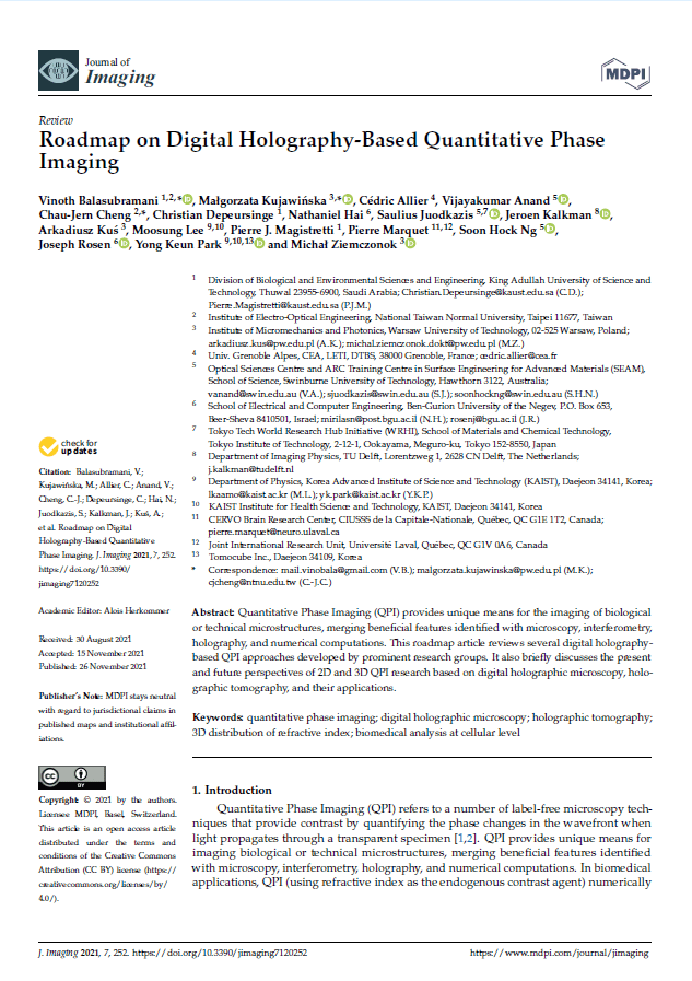
Roadmap on Digital Holography-Based Quantitative Phase Imaging
V. Balasubramani et al., Journal of Imaging, vol. 7, Art. no. 12, 2021
From digital holographic microscopy to optical coherence tomography – separate past and a common goal
A. Kuś, W. Krauze, and M. Kujawińska, Photonics Letters of Poland, vol. 13, Art. no. 4, 2021
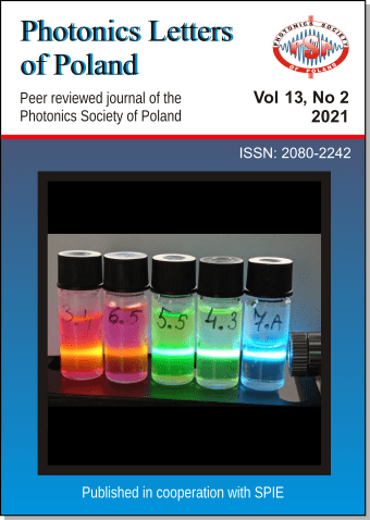
Reconstruction enhancement via projection screening in holographic tomography
P. Machnio, M. Ziemczonok, and M. Kujawińska, Photonics Letters of Poland, vol. 13, Art. no. 2, 2021
Enhanced QPI functionality by combining OCT and ODT methods
W. Krauze, P. Ossowski, M. Nowakowski, M. Szkulmowski, and M. Kujawińska, Quantitative Phase Imaging VII, 2021, vol. 11653, pp. 1–6
2020

Refractive Index Changes of Cells and Cellular Compartments Upon Paraformaldehyde Fixation Acquired by Tomographic Phase Microscopy
M. Baczewska, K. Eder, S. Ketelhut, B. Kemper, and M. Kujawińska, Cytometry Part A, vol. 97, Art. no. 9, 2020
Optical diffraction tomography with finite object support for the minimization of missing cone artifacts
W. Krauze, Biomedical Optics Express, vol. 11, Art. no. 4, 2020
Multi-incidence digital holographic microscopy with high axial resolution and enlarge measurement range
J. Martinez-Carranza, M. Mikuła-Zdańkowska, M. Ziemczonok, and T. Kozacki, Optics Express, vol. 28, Art. no. 6, 2020
Comparison of fixed and living biological cells parameters investigated with digital holographic microscope
P. K. Nienałtowski, M. Baczewska, and M. Kujawińska, Photonics Letters of Poland, vol. 12, Art. no. 1, 2020

Off-axis digital holographic multiplexing for rapid wavefront acquisition and processing
N. T. Shaked, M. Vicente, M. Trusiak, A. Kuś, and S. K. Mirsky, Advances in Optics and Photonics, vol. 12, Art. no. 3, 2020
Spatial bandwidth-optimized compression of image plane off-axis holograms with image and video codecs
P. Stępień, R. K. Muhamad, D. Blinder, P. Schelkens, and M. Kujawińska, Optics Express, vol. 28, Art. no. 19, 2020
Holotomographic investigation of an influence of PFA cell fixation process on refractive index of cellular organelles in epithelial cells
M. Baczewska, K. Eder, S. Ketelhut, B. Kemper, and M. Kujawińska, Quantitative Phase Imaging VI, 2020, vol. 11249, pp. 1–7
True Volumetric Measurements of Cells and Tissues by Limited Angle Holographic Tomography
M. Kujawińska and W. Krauze, Computational Optical Sensing and Imaging. Conference Proceedings 2020

Limited-angle holographic tomography for flow cytometry
A. Kuś, Digital Holography and Three-Dimensional Imaging 2020
Holographic data compression for holographic microscopy and tomography in biomedical applications
R. K. Muhamad, P. Stępień, D. Blinder, P. Schelkens, and M. Kujawińska, Digital Holography and Three-Dimensional Imaging 2020, pp. 1–2
Hologram compression in quantitative phase imaging
P. Stępień, R. K. Muhamad, M. Kujawińska, and P. Schelkens, Quantitative Phase Imaging VI, 2020, vol. 11249, pp. 1–12
Quantifying the performance of holographic tomography systems using the 3D-printed biological cell phantom
M. Ziemczonok, A. Kuś, and M. Kujawińska, Quantitative Phase Imaging VI, 2020, vol. 11249, pp. 1–7
2019

Polymer optical bridges for efficient splicing of optical fibers
M. Dudek and M. Kujawińska, Optical Engineering, vol. 58, Art. no. 2, 2019

Special Issue on Digital Holographic 3D Imaging: Capture, Display, and Evaluation
M. Kujawińska, B. Lee, J. Kim, and T. Kim, ETRI Journal, vol. 41, Art. no. 1, 2019
Holographic tomography: hardware and software solutions for 3D quantitative biomedical imaging
A. Kuś, W. Krauze, P. L. Makowski, and M. Kujawińska, ETRI Journal, vol. 41, Art. no. 1, 2019
Projection extrapolation routine for tight-frame limited-angle optical diffraction tomography
P. L. Makowski and M. Ziemczonok, Optics Letters, vol. 44, Art. no. 14, 2019
Digital Holographic Microscopy with extended field of view using tool for generic image stitching
P. Stępień, D. Korbuszewski, and M. Kujawińska, ETRI Journal, vol. 41, Art. no. 1, 2019

A "water window" tomography based on a laser-plasma double-stream gas-puff target soft X-ray source
P. W. Wachulak et al., Applied Physics B — Lasers and Optics, vol. 125, Art. no. 70, 2019
3D-printed biological cell phantom for testing 3D quantitative phase imaging systems
M. Ziemczonok, A. Kuś, P. Wasylczyk, and M. Kujawińska, Scientific Reports, vol. 9, pp. 1–9, 2019
Comparative study of laboratory and commercial limited-angle holographic tomography setups
M. Kujawińska, W. Krauze, M. Baczewska, A. Kuś, and M. Ziemczonok, Proceedings of SPIE: Quantitative Phase Imaging V, 2019, vol. 10887, pp. 1–7
Metrological studies of limited angle holographic tomography systems based on a phase phantom mimicking biological cell
M. Kujawińska, M. Ziemczonok, A. Kuś, and W. Krauze, Digital Holography and Three-Dimensional Imaging 2019, pp. 1–2
Projection multiplexing for enhanced acquisition speed in holographic tomography
A. Kuś, M. Baczewska, M. Ziemczonok, and M. Kujawińska, Three-Dimensional and Multidimensional Microscopy: Image Acquisition and Processing XXVI, Proceedings of SPIE, 2019, vol. 10883, pp. 1–6
Block-matching-based filtration in holographic tomography reconstruction
P. Stępień and M. Kujawińska, Proceedings of SPIE: Optical Methods for Inspection, Characterization, and Imaging of Biomaterials IV, 2019, vol. 11060, pp. 1–8
Tomographic imaging with the use of a compact soft X-ray microscope based on a laser plasma light source
P. Wachulak et al., EUV and X-ray Optics: Synergy between Laboratory and Space VI, 2019, vol. 11032, pp. 1–7
2018
Optonumerical method for improving functional parameters of polymer microtips
M. Dudek and M. Kujawińska, Optical Engineering, vol. 57, Art. no. 1, 2018

Reconstruction method for extended depth-of-field optical diffraction tomography
W. Krauze, A. Kuś, D. Śladowski, E. Skrzypek, and M. Kujawińska, Methods, vol. 136, pp. 40–49, 2018
Feasibility study of investigation of skin at cellular level by digital holographic microscopy
M. Baczewska, M. Kujawińska, E. Skrzypek, and D. Śladowski, Speckle 2018: VII International Conference on Speckle Metrology, 2018, vol. 10834, pp. 1–7
Polymer optical bridges for efficient splicing of optical fibers
M. Dudek and M. Kujawińska, Photonic Fiber and Crystal Devices: Advances in Materials and Innovations in Device Applications XII, 2018, vol. 10755, pp. 1–13
Sinogram cleaning procedure for optical diffraction tomography
W. Krauze and M. Kujawińska, Speckle 2018: VII International Conference on Speckle Metrology, 2018, vol. 10834, pp. 1–5
Introduction
M. Kujawińska and L. R. Jaroszewicz, Speckle 2018: VII International Conference on Speckle Metrology, 2018, vol. 10834, p. 1–1
Scanning errors in holographic tomography
A. Kuś, S. Mues, and B. Kemper, Speckle 2018: VII International Conference on Speckle Metrology, 2018, vol. 10834, pp. 1–6
Redundant Haar wavelet regularization in sparse-view optical diffraction tomography of microbiological structures
P. L. Makowski, Speckle 2018: VII International Conference on Speckle Metrology, 2018, vol. 10834, pp. 1–15
Multimodalne badania CKO-IR strukturalnych kompozytów polimerowych o strukturze kanapkowej poddanych działaniu wysokoenergetycznej wiązki laserowej
K. Siedlecki, M. Kujawińska, and P. K. Nienałtowski, Speckle 2018: VII International Conference on Speckle Metrology, 2018, vol. 10834, pp. 1–8
Multi-modal quantitative analysis of HeLa cells using digital holographic microscopy and confocal laser scanning microscopy
P. Stępień, T. Bernaś, W. Krauze, H. Sas-Nowosielska, and M. Kujawińska, Speckle 2018: VII International Conference on Speckle Metrology, 2018, vol. 10834, pp. 1–7
Characterization of 3D phantom for holographic tomography produced by two-photon polymerization
M. Ziemczonok, A. Kuś, M. Nawrot, and M. Kujawińska, Speckle 2018: VII International Conference on Speckle Metrology, 2018, vol. 10834, pp. 1–7
2017
Illumination-related errors in limited-angle optical diffraction tomography
A. Kuś, Applied Optics, vol. 56, Art. no. 33, 2017
Phase retrieval with tunable phase transfer function based on the transport of intensity equation
J. Martinez-Carranza, P. Stępień, and T. Kozacki, Modeling Aspects in Optical Metrology VI, Proceedings Volume 10330, 2017, pp. 1–10
Reconstruction method for extended depth-of-field limited-angle tomography
W. Krauze, A. Kuś, E. Skrzypek, and M. Kujawińska, Quantitative Phase Imaging III, 2017, vol. 10074, pp. 1–5
Focus-tunable lens in limited-angle holographic tomography
A. Kuś, W. Krauze, and M. Kujawińska, Three-Dimensional and Multidimensional Microscopy: Image Acquisition and Processing XXIV, Proceedings of SPIE, 2017, vol. 10070, pp. 1–9